")
")
Support for the case study of invasive maxillary cyst in collaboration with the dental specialist Dr. Michele Di Cosola at the Villa Pompea, excellence facility in Bari.
How is innovation improving the working approach and traditional methodologies for the treatment of complex cases?
In recent years we are noticing how 3D technologies are entering the medical sector forcefully. The innovative techniques greatly improve the work of the specialists in the field, offering ever faster and more efficient solutions. Surgeon specialists are almost always faced with new and complex case studies, which represent real challenges every time they cross the threshold of the operating room.
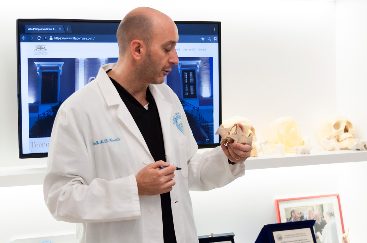
Dr. Michele Di Cosola
For the medical operator surgical planning represents a key phase that can significantly lighten, in terms of timing and fatigue, the decisive phase of direct action on the anatomical district. Very often the analysis and the clinical case study carried out on CT images before surgery are not enough, since they offer a limited 2D view.
The overview of the district and the details on the location as size of a lesion are limited. Often during the direct action on the area and on the tissue the operator may be faced with an unexpected situation far from his predictions, with consequences that could cause discomfort for the patient and the surgeon himself, with the lengthening of the intervention times.
3D technologies can greatly improve all pre/post-operative phases, bringing benefits and advantages that are increasingly appreciated.
The use of increasingly innovative and sector-specific tools has simplified the methods of approach to the pre-operative preparation. The surgeon, thanks to the 3D physical model, manages to put the patient at his ease, explaining the procedures adopted in the operating room as well as planning all the access and treatment steps under examination. The entire daily workflow within the medical facility will be smoother and optimized, thanks to the reduction of the O.R. employment time.
The attention to detail guaranteed by 3D printing is particularly necessary in surgical areas, such as the maxillofacial sector.
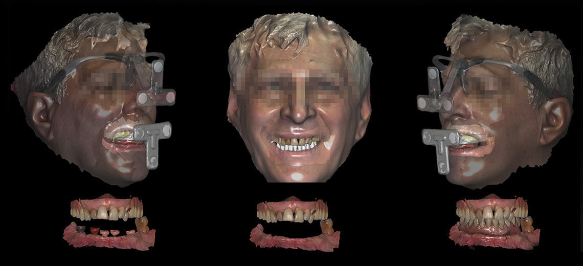
The CreaMED team worked on a case study in collaboration with Dr. Di Cosola, maxillofacial surgeon and renowned specialist in the sector: patient with maxillary cyst, unique and specific case, for which the doctor has chosen to plan an operative treatment studied in detail, as the anatomical area concerned is particularly delicate.
The case required a careful evaluation of the anatomical areas. Di Cosola explains:
"The patient has a voluminous right maxillary cyst with an invasive and obstructing attitude. This lesion is of apical-radicular source, originating from the apex of some teeth, in particular from the right premolars that generated this expansive growth lesion."
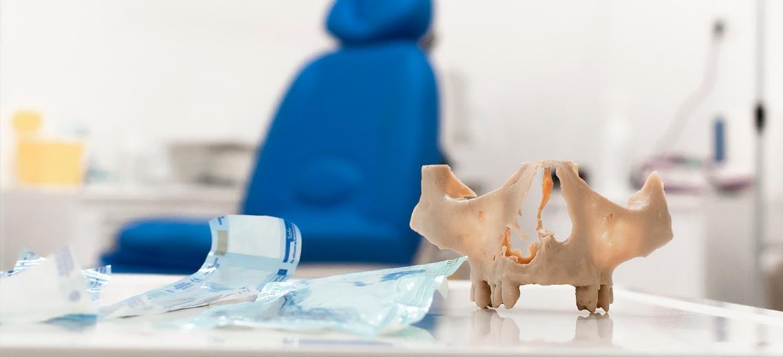
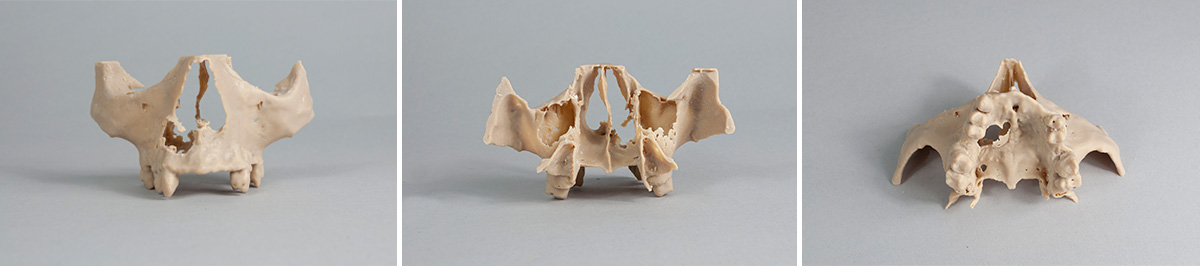
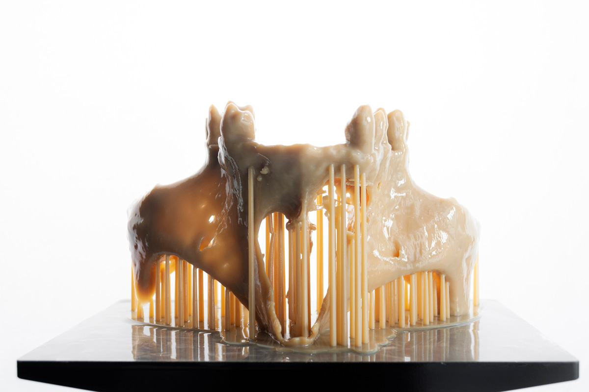
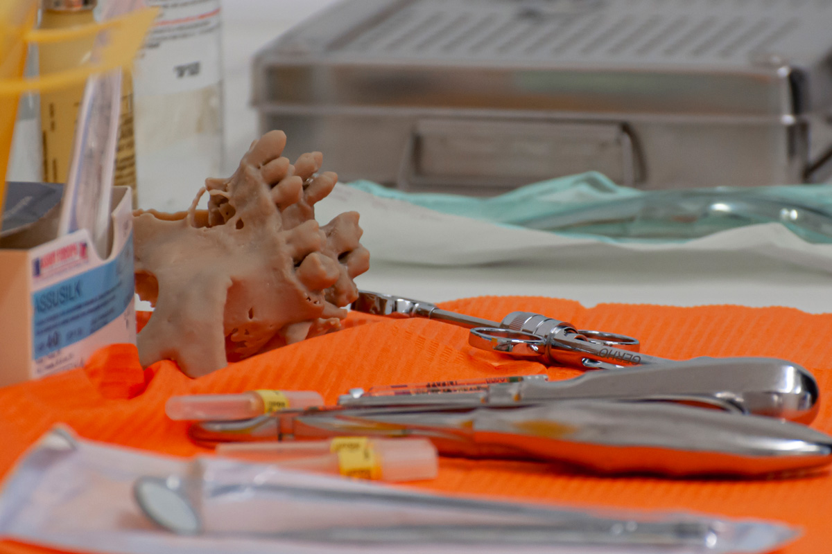
Thanks to specific software designed for the reconstruction of anatomical areas from CT images, with algorithms based on image segmentation, we focused on the maxillary and palate bone area affected by the lesion. Once the virtual 3D model was obtained, it was prepared for 3D printing, choosing a resin printing technology (DLP) that guarantees safety in terms of precision up to the cent.
The surgeon particularly appreciated the fidelity in the anatomical details of the model up to the bone residues of the maxillary cortical area eroded by the cyst. Before entering the room, the doctor reassured the patient by explaining the anatomical complication and the surgical procedure adopted, with the help of the printed model.
By identifying the condition and size of the lesion on the model, the doctor was able to identify the flow of intervention, explaining:
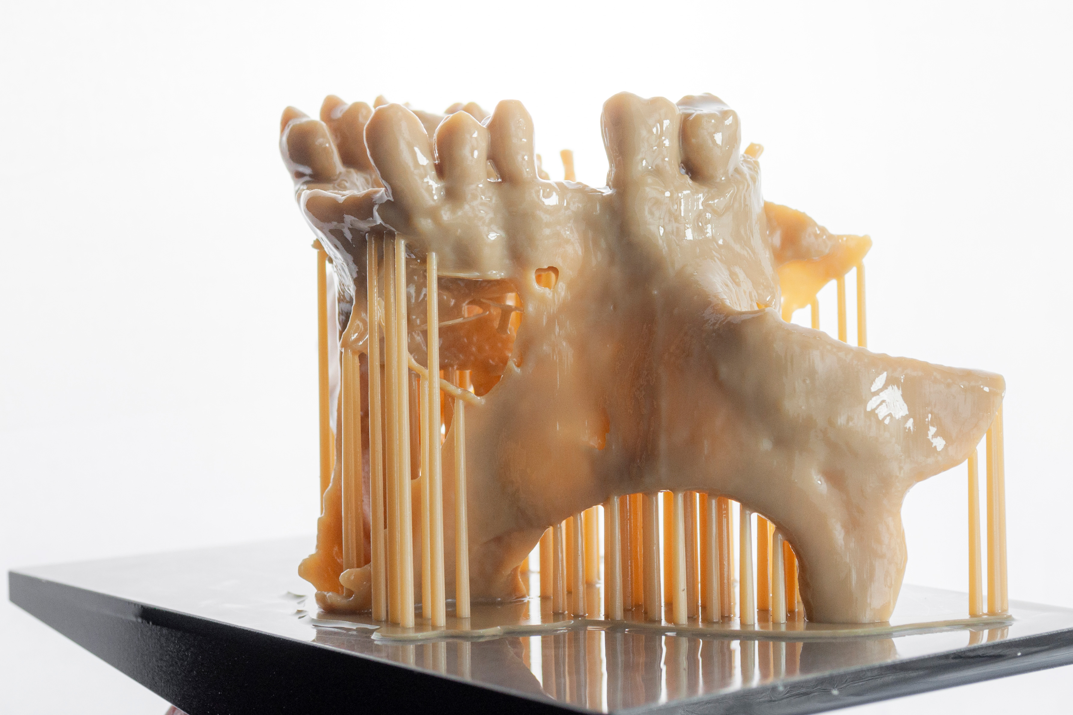
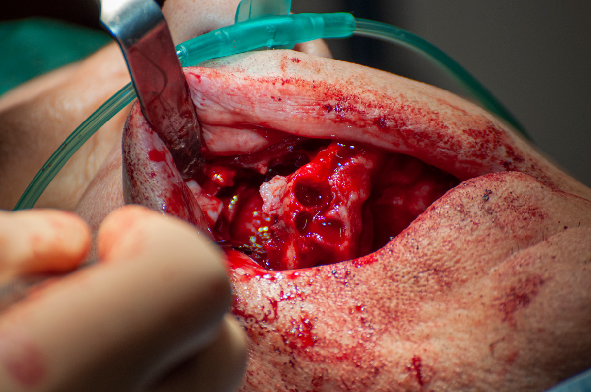
“During its evolutionary process this lesion has eroded the bone component of the patient's skeletal structure, so in its anatomical dissection, it must be borne in mind that it is important to find a cleavage plane within the maxillary sinus. I chose to use the 3D method to build a virtual model that would bring the patient's natural anatomy to a 1:1 scale, to understand the limits of the lesion, and to be able to remove it completely without creating further damage to the patient."
"Surely, judging from the clinical conditions, this lesion also infiltrates soft tissues such as the floor of the nose, from the nasal mucosa to the orbicularis muscle of the mouth which is inserted on the external face of the maxilla as well as having an extension up to the limit with the Bichat. "
Finally, Di Cosola highlighted the precision characteristics obtained on the model:
“In the reconstructed three-dimensional model, we can appreciate small bony segments that continue to represent the lower edge of the nose. We can see, using such a precise three-dimensional model, how this lesion has also eroded part of the palate, we expect how from the palate we can get to the nasal cavity. During the removal of the lesion, one must be careful not to have an invasion of the nasal soft tissue structures and free it of all the residues inserted on the floor of the maxillary sinus which has now been eroded.”
The surgeon's choice to introduce 3D printing to support the analysis and planning of the intervention situation finally proved effective and ameliorative for him. The benefits brought by the use of these technologies continue to improve in relation to the continuous development of additive technologies.